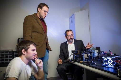
Ulas Sunar, right, Ohio Research Scholar for Medical Imaging and Biomedical Imaging Laboratory director, is developing an endoscope to detect and treat cancer. He is pictured with researchers Jeremy Kress, left, and Dan Rohrbach. (Photo by Erin Pence)
Developing a revolutionary technology to detect and treat cancerous tumors in the intraperitoneal cavity and other internal cavities such as oral and gastrointestinal is the goal of a research project being led by Wright State University physicist Ulas Sunar.
Sunar is an Ohio Research Scholar for Medical Imaging, new director of the Biomedical Imaging Laboratory and associate professor in the College of Engineering and Computer Science’s Department of Biomedical, Industrial and Human Factors Engineering and the College of Science and Mathematics.
In the Biomedical Imaging Lab in the new Neuroscience Engineering Collaboration Building, Sunar and his research team are building a new kind of flexible, fiber-based endoscope that is to be inserted into a body cavity or organ. It is a part of a two-year, $412,093 grant from the National Institute of Biomedical Imaging and Bioengineering division of the National Institutes of Health.
Sunar plans to increase the sensitivity of the endoscope by adding a quantitative fluorescent imaging component, which can better detect lesions on the surface of internal organs. Blood and tissue tends to absorb conventional light and diminish the fluorescence imaging contrast. But the new fluorescent endoscope would correct for that, resulting in an improved contrast so that clinicians can better delineate cancer from surrounding normal tissue.
There is a therapy component to the approach, Sunar says. Surgeons often destroy cancer cells using a fixed laser beam pattern, but lesions usually have irregular shapes.
“What we do in this endoscope is that we project a laser light beam that is customized to the shape of the tumor,” said Sunar. “In that way, all of the cancerous cells are removed, but the healthy cells remain intact.”
A novel chemo-drug is being developed by researchers at the University of Buffalo in which a cancer-killing drug in nanoparticles encapsulated by liposomes is injected into the patient and is then activated by the endoscope Sunar is developing.
“We shine the light and it triggers the release of the drug only in the targeted tumor area,” said Sunar. “The endoscopic device has an imaging and a therapy component. It’s a novel idea.”
Once Sunar and his research team complete the endoscope, they will begin testing it on an animal model.
He said the NIH grant has the potential to be extended to up to five years that would include clinical studies.

 Wright State psychology team studies ways to identify fatigue in pilots, drivers
  Wright State videographer Kris Sproles wins Regional Emmy and Ohio journalism award
  Wright State Boonshoft School of Medicine ranked among the nation’s best for 2024 by U.S. News
  Exposing biotechnology
  Wright State faculty member Dan Noel uses unique background to inspire new leaders